[1] Liu R, Yang J, Yao J, et al. Optogenetic control of RNA function and metabolism using engineered light-switchable RNA-binding proteins. Nat Biotechnol. 2022;40(5):779-786. doi:10.1038/s41587-021-01112-1(IF=68.164)
[2] Luo J, Yang Q, Zhang X, et al. TFPI is a colonic crypt receptor for TcdB from hypervirulent clade 2 C. difficile. Cell. 2022;185(6):980-994.e15. doi:10.1016/j.cell.2022.02.010 (IF=66.85)
[3] Zhou J, Chen P, Wang H, et al. Cas12a variants designed for lower genome-wide off-target effect through stringent PAM recognition. Mol Ther. 2022;30(1):244-255. doi:10.1016/j.ymthe. 2021.10.010 (IF=12.910)
[4] Chen S, Cao X, Zhang J, Wu W, Zhang B, Zhao F. circVAMP3 Drives CAPRIN1 Phase Separation and Inhibits Hepatocellular Carcinoma by Suppressing c-Myc Translation. Adv Sci (Weinh). 2022;9(8):e2103817. doi:10.1002/advs.202103817 (IF=17.694)
[5] Zhang Y, Yu X, Sun R, et al. Splicing factor arginine/serine-rich 8 promotes multiple myeloma malignancy and bone lesion through alternative splicing of CACYBP and exosome-based cellular communication. Clin Transl Med. 2022;12(2):e684. doi:10.1002/ctm2.684 (IF=11.492)
[6] Qin J, Cai Y, Xu Z, et al. Molecular mechanism of agonism and inverse agonism in ghrelin receptor. Nat Commun. 2022;13(1):300. Published 2022 Jan 13. doi:10.1038/s41467-022-27975-9 (IF=17.681)
[7] Tang X, Deng Z, Ding P, et al. A novel protein encoded by circHNRNPU promotes multiple myeloma progression by regulating the bone marrow microenvironment and alternative splicing. J Exp Clin Cancer Res. 2022;41(1):85. Published 2022 Mar 8. doi:10.1186/s13046-022-02276-7(IF=12.658)
[8] Yang X, Wang X, Xu Z, et al. Molecular mechanism of allosteric modulation for the cannabinoid receptor CB1 [published online ahead of print, 2022 May 30]. Nat Chem Biol. 2022;10.1038/s41589-022-01038-y. doi:10.1038/s41589-022-01038-y (IF=16.174)
[9] Xie F, Su P, Pan T, et al. Engineering Extracellular Vesicles Enriched with Palmitoylated ACE2 as COVID-19 Therapy. Adv Mater. 2021;33(49):e2103471. doi:10.1002/adma. 202103471 (IF=30.849)
[10] Liang Y, Lu Q, Li W, et al. Reactivation of tumour suppressor in breast cancer by enhancer switching through NamiRNA network. Nucleic Acids Res. 2021;49(15):8556-8572. doi:10.1093/nar/gkab626 (IF=16.9)
[11] Fan Y, Wang J, Jin W, et al. CircNR3C2 promotes HRD1-mediated tumor-suppressive effect via sponging miR-513a-3p in triple-negative breast cancer. Mol Cancer. 2021;20(1):25. Published 2021 Feb 2. doi:10.1186/s12943-021-01321-x (IF=27.403)
[12] Dai L, Dai Y, Han J, et al. Structural insight into BRCA1-BARD1 complex recruitment to damaged chromatin. Mol Cell. 2021;81(13):2765-2777.e6. doi:10.1016/j.molcel.2021.05.010 (IF=17.97)
[13] Zhang K, Wang A, Zhong K, et al. UBQLN2-HSP70 axis reduces poly-Gly-Ala aggregates and alleviates behavioral defects in the C9ORF72 animal model. Neuron. 2021;109(12):1949-1962.e6. doi:10.1016/j.neuron.2021.04.023 (IF=17.17)
[14] Liang Y, Lu Q, Li W, et al. Reactivation of tumour suppressor in breast cancer by enhancer switching through NamiRNA network. Nucleic Acids Res. 2021;49(15):8556-8572. doi:10.1093/nar/gkab626 (IF=16.9)
[15] Li T, Chen X, Qian Y, et al. A synthetic BRET-based optogenetic device for pulsatile transgene expression enabling glucose homeostasis in mice. Nat Commun. 2021;12(1):615. Published 2021 Jan 27. doi:10.1038/s41467-021-20913-1 (IF=14.92)
[17] Gu C, Wang Y, Zhang L, et al. AHSA1 is a promising therapeutic target for cellular proliferation and proteasome inhibitor resistance in multiple myeloma. J Exp Clin Cancer Res. 2022;41(1):11. Published 2022 Jan 6. doi:10.1186/s13046-021-02220-1 (IF=11.161)
[18] Zhou Y, Li D, Luo J, et al. Sulfated glycosaminoglycans and low-density lipoprotein receptor mediate the cellular entry of Clostridium novyi alpha-toxin. Cell Res. 2021;31(8):935-938. doi:10.1038/s41422-021-00510-z (IF=25.617)
[19] Luo Q, Wu X, Zhao P, et al. OTUD1 Activates Caspase-Independent and Caspase-Dependent Apoptosis by Promoting AIF Nuclear Translocation and MCL1 Degradation. Adv Sci (Weinh). 2021;8(8):2002874. Published 2021 Feb 8. doi:10.1002/advs.202002874 (IF=15.84)
[20] Yan F, Huang C, Wang X, et al. Threonine ADP-Ribosylation of Ubiquitin by a Bacterial Effector Family Blocks Host Ubiquitination. Mol Cell. 2020;78(4):641-652.e9. doi:10.1016/j.molcel.2020.03.016 (IF=17.97)
[21] Sun X, Peng X, Cao Y, Zhou Y, Sun Y. ADNP promotes neural differentiation by modulating Wnt/β-catenin signaling. Nat Commun. 2020;11(1):2984. Published 2020 Jun 12. doi:10.1038/s41467-020-16799-0 (IF=14.911)
[22] Yang X, Wang H, Xie E, et al. Rewiring ERBB3 and ERK signaling confers resistance to FGFR1 inhibition in gastrointestinal cancer harbored an ERBB3-E928G mutation. Protein Cell. 2020;11(12):915-920. doi:10.1007/s13238-020-00749-z (IF=14.872)
[23] Zou Y, Wang A, Shi M, et al. Analysis of redox landscapes and dynamics in living cells and in vivo using genetically encoded fluorescent sensors. Nat Protoc. 2018;13(10):2362-2386. doi:10.1038/s41596-018-0042-5 (IF=13.490)
[24] Hao H, Hu S, Chen H, et al. Loss of Endothelial CXCR7 Impairs Vascular Homeostasis and Cardiac Remodeling After Myocardial Infarction: Implications for Cardiovascular Drug Discovery. Circulation. 2017;135(13):1253-1264. doi:10.1161/CIRCULATIONAHA.116.023027 (IF=18.881)


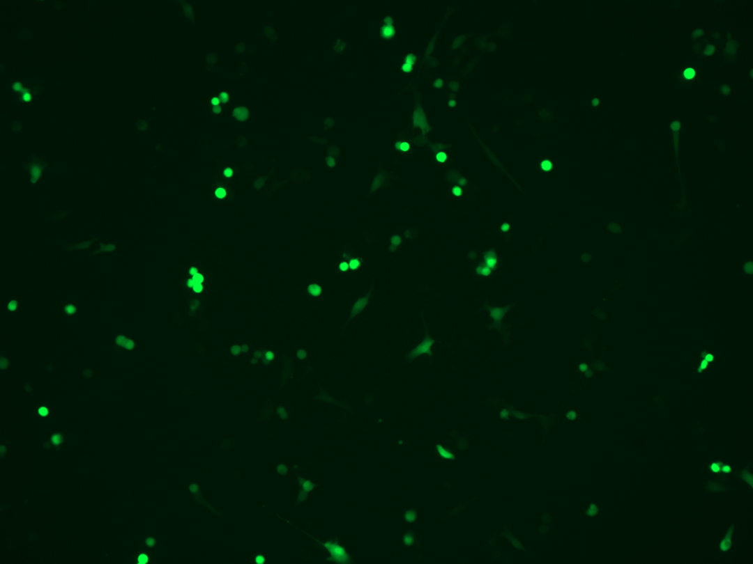
 细胞转染实验失败了,怎么解救?
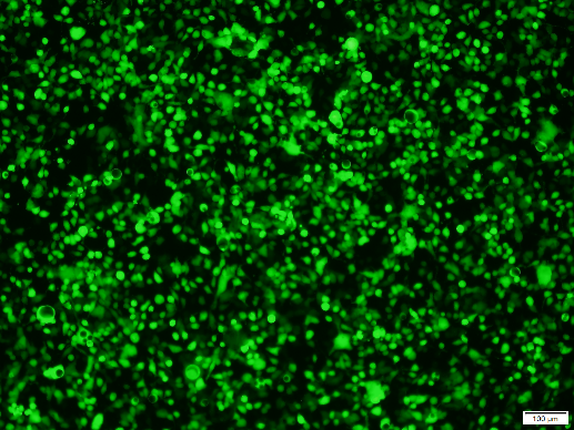